
Dysembryoplastic Neuroepithelial Tumor with Atypical Presentation: MRI and Diffusion Tensor Characteristics
DOI:
https://doi.org/10.3941/jrcr.v7i11.1559Keywords:
Dysembryoplastic neuroepithelial tumor (DNET), Diffusion tensor imaging, Neuroimaging, TractographyAbstract
We report the neuroimaging findings of a 26-year-old female patient with a biopsy-proven dysembryoplastic neuroepithelial tumor (DNET). DNETs are an uncommon, usually benign, glial-neural cortical neoplasm of children and young adults who typically present with intractable seizures. DNETs may occur in any region of the supratentorial cortex, but have a predilection for the temporal lobes. Accurate neuroimaging diagnosis is essential since patients with DNET benefit from complete resection. However, accurate differentiation from other cortical lesions may be challenging. Typical conventional Magnetic Resonance Imaging (MRI) features can help in the differentiation from other similar cortical tumors. Diffusion tensor imaging can also provide important additional diagnostic information regarding the degree of involvement of adjacent parenchyma and white matter tracts. In this case, tractography and fractional anisotropy maps demonstrated that fiber tracts surrounding the lesion were displaced, but fiber integrity was maintained, which is more suggestive of a DNET rather than a more aggressive neoplasm. Accurate identification of DNETs is essential for the purpose of rendering a timely diagnosis and start appropriate treatment.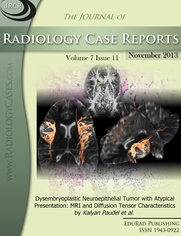
Downloads
Published
2013-11-26
Issue
Section
Neuroradiology
License
The publisher holds the copyright to the published articles and contents. However, the articles in this journal are open-access articles distributed under the terms of the Creative Commons Attribution-NonCommercial-NoDerivs 4.0 License, which permits reproduction and distribution, provided the original work is properly cited. The publisher and author have the right to use the text, images and other multimedia contents from the submitted work for further usage in affiliated programs. Commercial use and derivative works are not permitted, unless explicitly allowed by the publisher.


