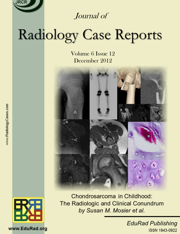
Chondrosarcoma in Childhood: The Radiologic and Clinical Conundrum
DOI:
https://doi.org/10.3941/jrcr.v6i12.1241Keywords:
pediatric chondrosarcoma, chondrosarcoma, bone tumor, pediatricAbstract
Less than 10% of chondrosarcomas occur in children. In addition, as little as 0.5% of low-grade chondrosarcomas arise secondarily from benign chondroid lesions. The presence of focal pain is often used to crudely distinguish a chondrosarcoma (which is usually managed with wide surgical excision), from a benign chondroid lesion (which can be followed by clinical exams and imaging surveillance). Given the difficulty of localizing pain in the pediatric population, initial radiology findings and short-interval follow-up, both imaging and clinical, are critical to accurately differentiate a chondrosarcoma from a benign chondroid lesion. To our knowledge, no case in the literature discusses a chondrosarcoma possibly arising secondarily from an enchondroma in a pediatric patient. We present a clinicopathologic and radiology review of conventional chondrosarcomas. We also attempt to further the understanding of how to manage a chondroid lesion in the pediatric patient with only vague or bilateral complaints of pain.Downloads
Published
2012-12-26
Issue
Section
Pediatric Radiology
License
The publisher holds the copyright to the published articles and contents. However, the articles in this journal are open-access articles distributed under the terms of the Creative Commons Attribution-NonCommercial-NoDerivs 4.0 License, which permits reproduction and distribution, provided the original work is properly cited. The publisher and author have the right to use the text, images and other multimedia contents from the submitted work for further usage in affiliated programs. Commercial use and derivative works are not permitted, unless explicitly allowed by the publisher.


