
Diagnostic dilemma in a neglected case of fetus-in-fetu solved with Magnetic Resonance Imaging and MDCT - a case report and review of literature
DOI:
https://doi.org/10.3941/jrcr.v5i10.833Keywords:
fetus-in-fetu, teratoma, axial skeleton, diamniotic monochorionic twins, parasitic twin, multidetector computed tomography, magnetic resonance imagingAbstract
Fetus-in-fetu (FIF) is a rare anomaly in which a vertebrate fetus is enclosed within the body of its twin in diamniotic monochorionic pregnancy. To the best of our knowledge, fewer than 100 cases have been reported in literature. Although a wide variety of presentations have been described in clinical reports, the characteristic features on MRI which distinguish FIF from teratoma have not been well delineated. Here we present a case of fetus-in-fetu in which characteristic MDCT and MR findings were used to diagnose FIF preoperatively and successfully differentiate it from teratoma. Although both CT and MRI can be used for definitive preoperative diagnosis of FIF, MRI is an ideal imaging modality due to inherent high tissue contrast and spatial resolution. Furthermore, MRI obviates the need for iodine contrast and eliminates the risk of ionizing radiation. We emphasize that MRI is an ideal valuable diagnostic tool for definite preoperative diagnosis of FIF and surgical planning.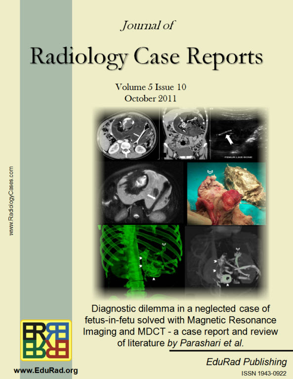
Downloads
Published
2011-10-09
Issue
Section
Pediatric Radiology
License
The publisher holds the copyright to the published articles and contents. However, the articles in this journal are open-access articles distributed under the terms of the Creative Commons Attribution-NonCommercial-NoDerivs 4.0 License, which permits reproduction and distribution, provided the original work is properly cited. The publisher and author have the right to use the text, images and other multimedia contents from the submitted work for further usage in affiliated programs. Commercial use and derivative works are not permitted, unless explicitly allowed by the publisher.


