
Bilateral Thalamic and Right Fronto-temporo-parietal Gliomas in a 4 Years Old Child Diagnosed by Magnetic Resonance Imaging
DOI:
https://doi.org/10.3941/jrcr.v10i1.2306Keywords:
Bithalamic gliomas, cortical gliomas, intracranial mass, paediatric seizures, magnetic resonance imagingAbstract
We report the neuroimaging findings of a 4-year-old girl with biopsy-proven bilateral thalamic and right fronto-temporo-parietal cortical gliomas, which are uncommon tumours involving the central nervous system. Despite their benignity, the prognosis is usually poor because of involvement of the thalamic nuclei and difficulty in surgical excision. These lesions have limited differential diagnoses that include metabolic, toxic, infective, vascular and neoplastic. Imaging characteristics on conventional Magnetic Resonance (MR), Magnetic Resonance Spectroscopy (MRS) and Diffusion tensor imaging (DTI) can further narrow the differential diagnosis and also provide additional information regarding the degree of involvement of adjacent brain tissue and white matter tracts around the lesions.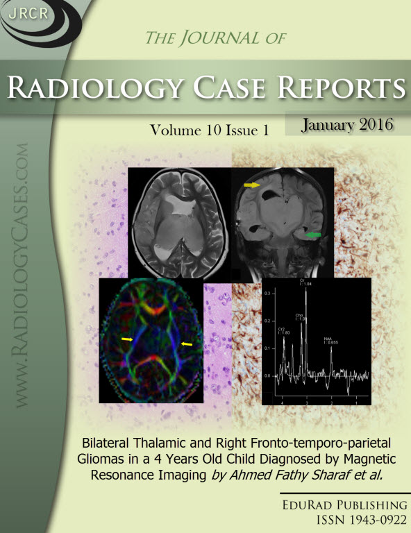
Downloads
Published
2016-01-27
Issue
Section
Neuroradiology
License
The publisher holds the copyright to the published articles and contents. However, the articles in this journal are open-access articles distributed under the terms of the Creative Commons Attribution-NonCommercial-NoDerivs 4.0 License, which permits reproduction and distribution, provided the original work is properly cited. The publisher and author have the right to use the text, images and other multimedia contents from the submitted work for further usage in affiliated programs. Commercial use and derivative works are not permitted, unless explicitly allowed by the publisher.


